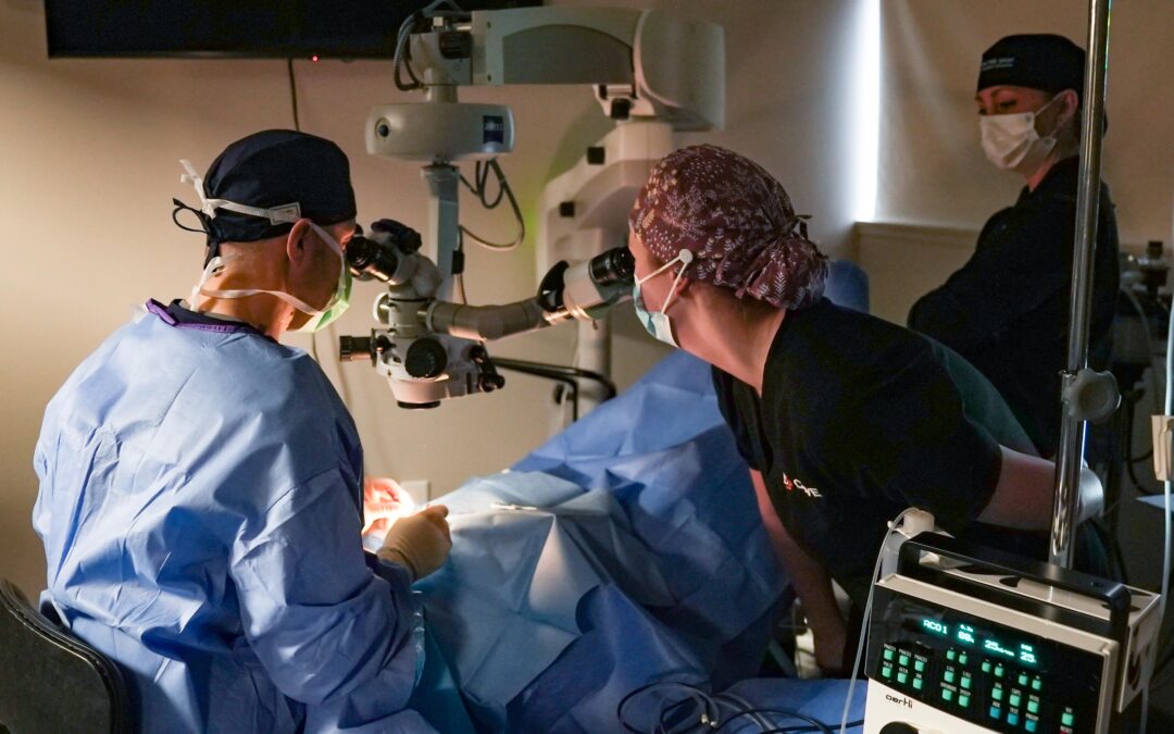
Eric J. Miller, DVM, MS, Diplomate ACVO recently performed a successful Cataract surgery at our affiliate COVE – Veterinary Specialty & Emergency Hospital.
The surgery was on a diabetic dog that went pretty acutely blind with cataracts. Diabetes in dogs causes cataracts in virtually all dogs and most of them go blind within 6 months of their diagnosis of diabetes. Cataract surgery (phacoemulsification) is currently the only way to restore vision in these cases. Our patient had become diabetic while the client was in Arizona help her daughter recover from a surgery and caring for her daughters’ animals. They didn’t figure everything out until they got back from Arizona and Apricot was blind. After some testing to make sure she was a good candidate for surgery we went forward with the procedure. In the procedure we work through a microscope to remove the existing opaque lens cells through a process called phacoemulsification and maintain what is called the “lens capsule”. After the damaged cells are removed, we place a special made foldable acrylic intraocular lens into the existing “lens capsule” to restore lens functionality. The procedure in animals is for all intensive purposes the same as cataract surgery in humans and the equipment we use is not animal specific it is the same as the equipment used in people. The first artificial intraocular lens was developed and placed by a British ophthalmologist named Sir Harold Ridley (Knighted for his achievement) in 1949. Phacoemulsification is the modern approach to cataract removal and it was developed and introduced by American ophthalmologist Dr. Charles Kelman in 1967. The dog eye is quiet a bit bigger than a humans and the lens is about 3-4 times larger for comparisons sake.